Require Assistance?
Get A Quick Callback From Our Healthcare Experts
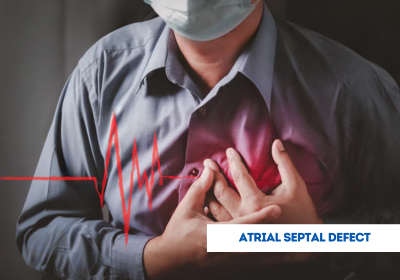
Atrial Septal Defect (ASD) Closure addresses a hole in the wall between the heart’s upper chambers, which allows oxygen-rich and oxygen-poor blood to mix. Large ASDs can strain the heart and lungs, leading to fatigue, breathing difficulties, or infections. Timely closure is crucial to prevent complications. With EdhaCare, patients can access atrial septal defect (ASD) closure in India, Turkey, Thailand, Dubai, and other leading countries at advanced cardiac centers with complete support and assistance throughout the treatment journey.
Individuals who have the following need ASD closure:
Infants and children are usually observed for spontaneous closure of the defect, but treatment is suggested when it does not close spontaneously by early childhood or when symptoms develop. Adults with undiagnosed ASD may require closure if significant complications develop.
The cost of atrial septal defect closure can fluctuate based on the procedure type (surgical or device closure), hospital facilities, complexity of the defect, and the expertise of the cardiologists or cardiothoracic surgeons performing the procedure. Below are approximate costs for atrial septal defect closure in India, Turkey, Thailand, and Dubai:
| Country | Approximate Cost |
|---|---|
| India | USD 6,000 – 20,000 |
| Turkey | USD 12,000 – 30,000 |
| Thailand | USD 15,000 – 35,000 |
| Dubai | USD 18,000 – 45,000 |
Note: Above costs for atrial septal defect closure (ASD) Closure are estimated. Reach out to EdhaCare for exact cost of atrial septal defect closure (ASD) Closure and personalized guidance.
Here are the top hospitals in India for atrial septal defect (ASD) closure. These best hospitals for atrial septal defect (ASD) closure are known for their modern cardiac facilities, advanced imaging technology, and experienced teams that focus on safely closing the heart defect and improving long-term heart health.
| Hospital | Location |
|---|---|
| Fortis Escorts Heart Institute | New Delhi |
| Apollo Children’s Hospital | Chennai |
| Max Super Specialty | Delhi |
| KIMS Hospital | Hyderabad |
| Artemis Hospital | Gurgaon |
Looking for top doctors in India for atrial septal defect (ASD) closure? EdhaCare connects you with the best doctors for ASD closure in India to ensure the most effective outcomes.
| Doctor | Hospital | Location |
|---|---|---|
| Dr. Rajesh Sharma | Jaypee Hospital | Noida |
| Dr. K R Balakrishnan | Fortis Malar Hospital | Chennai |
| Dr. Kulbhushan Singh Dagar | Max Super Specialty | Delhi |
| Dr. Sandeep Attawar | KIMS Hospital | Hyderabad |
| Dr. Devi Prasad Shetty | Narayana Health | Bangalore |
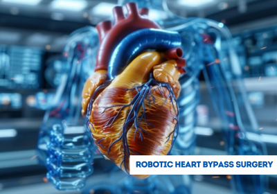
Selecting a procedure for ASD closure will depend not only on the size, location, and degree of severity but also on the type of defect. The two main types include:
Both types of repairs have excellent success rates. The catheter-based procedure typically allows faster recovery for patients and has less scarring associated with surgery.
Before ASD closure, a comprehensive heart examination consisting of the following is done.
A cardiologist and cardiothoracic team will use five factors to plan the best approach to treatment, namely.
When it comes to catheter-based closures, the anatomy should be responsive to the anchoring of the device. Growth potential and long-term outcomes are also discussed in children.
Catheter-Based Closure Procedure (Minimally Invasive):
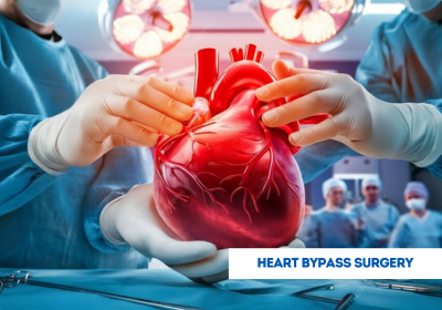
Surgical ASD Repair:
Although ASD repair is a safe and usual procedure, there are specific hazards:
Catheter-Based Closure Risks:
Surgical Closure Risks:
These two procedures are associated with low rates of complications and a great safety profile in experienced centers.
Post-Surgical Repair
Post-Catheter Procedure
Long-term care is necessary for guaranteeing heart health and tracking down any complications and may include the following:
Although a majority of patients return to normal life and children get back to sports after the cardiologist's clearance, some patients can develop rhythm disturbances in the future. So, ASD closure becomes a huge improvement in life expectancy and quality of life.
India boasts one of the best success rates for ASD closure thanks to the advanced interventional cardiology and pediatric cardiac surgery programs.
Indian hospitals report low complication rates with excellent results in infants and small children, too. Prompt diagnosis and closure eliminate the long-term effects, such as heart failure or stroke.
India is one of the most trusted countries to undergo ASD closure because of advanced technology, seasoned cardiologists, and low cost of care.
Key Benefits:
Hospitals such as Narayana Health and Apollo Hospitals employ real-time 3D echocardiography and hybrid cath labs for accurate ASD closures.
For international patients looking for ASD closure in India, it is necessary to present certain documentation to have a smooth medical journey. These include:
It is advisable to refer to the Indian consulate or your medical facilitator for the latest information and help in documentation.
Is a self-closing ASD possible?
Small ASDs, particularly in infants, can close without medical treatment. Larger defects usually require intervention.
Is ASD surgery risky?
ASD closure is safe with high success rates, especially when they are performed by experienced specialists.
Do I have to get a pacemaker after the closure of ASD?
Most patients do not, but there are the oddities that require one (rare cases with rhythm disturbances).
Can adults undergo ASD closure?
Indeed, adults who were not diagnosed with ASD before or suffer from its symptoms can receive tremendous help from closure.
How long do children need to wait before getting back to school or any activity after the closure of ASD?
Usually, within 2 to 3 weeks after the catheter-based closure and 4 to 6 weeks after the surgical repair.


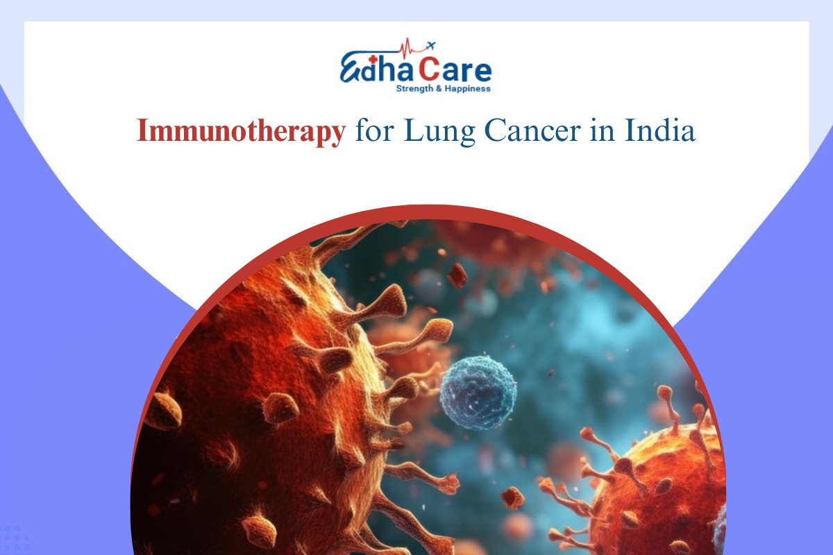
Lung cancer is the most commonly diagnosed cancer across the globe and it serves as the primary reas...

Are you planning to travel from the United Kingdom to India for your treatment but uncertain about t...
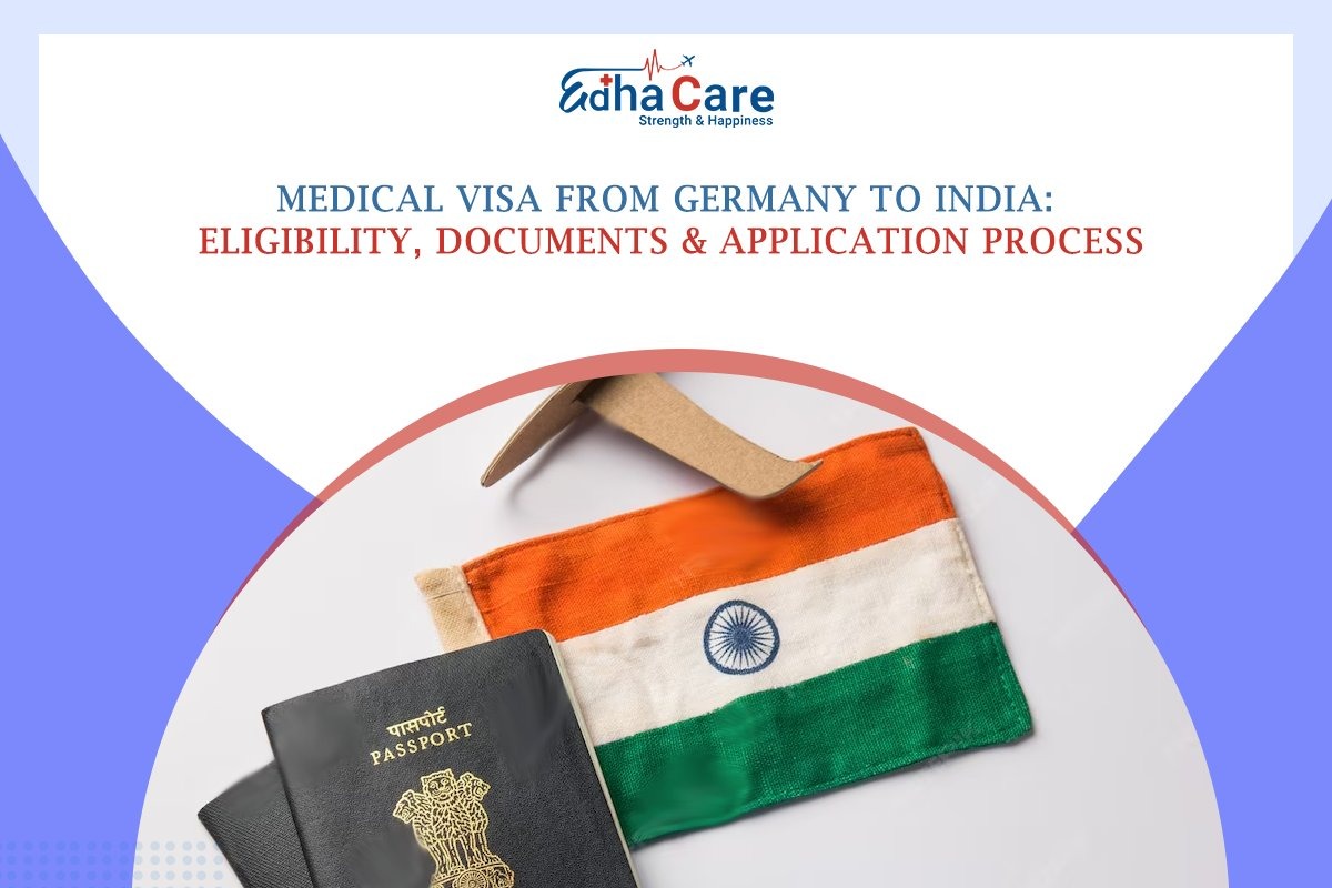
Planning to travel from Germany to India for medical treatment? Confused about the steps involved in...