Tetralogy Of Fallot
Tetralogy of Fallot (TOF) is a congenital heart defect characterised by a combination of four abnormalities in the heart's structure. Surgery is the primary treatment option for individuals with TOF to correct the underlying cardiac anomalies and restore normal blood flow. In this article, we will explore the concept of surgery for Tetralogy of Fallot, its significance, and the procedure involved in addressing this complex cardiac condition.
Book an AppointmentAbout Tetralogy Of Fallot
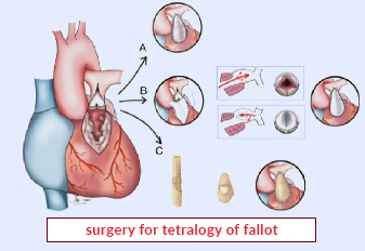
Tetralogy of Fallot is a complex congenital heart defect that involves four primary abnormalities:
-
Ventricular Septal Defect (VSD): A hole in the wall separating the heart's lower chambers (ventricles), allowing blood to mix between the two chambers.
-
Pulmonary Stenosis: Narrowing of the pulmonary valve or the blood vessel that carries blood from the heart to the lungs, restricting the flow of blood to the lungs for oxygenation.
-
Overriding Aorta: The aorta, the main blood vessel that carries oxygen-rich blood to the body, is positioned over both ventricles rather than emerging solely from the left ventricle.
-
Right Ventricular Hypertrophy: The right ventricle, responsible for pumping blood to the lungs, becomes thickened and enlarged due to the increased workload.
Procedure of Tetralogy Of Fallot
-
Preoperative Evaluation: Before surgery, a comprehensive evaluation is performed to assess the patient's overall health, cardiac function, and anatomical variations of Tetralogy of Fallot. This may include echocardiography, cardiac catheterization, and other imaging tests.
-
Anesthesia and Incision: Surgery for Tetralogy of Fallot Surgery is performed under general anaesthesia. An incision is made in the chest, typically through a median sternotomy (vertical incision through the breastbone), allowing access to the heart.
-
Cardiopulmonary Bypass: The patient is connected to a heart-lung bypass machine, which temporarily takes over the function of the heart and lungs during the surgery. The machine ensures blood circulation and oxygenation while the surgeon operates on the heart.
-
Surgical Repair: The specific surgical approach depends on the patient's individual anatomy and the severity of the cardiac abnormalities. The surgeon typically addresses the ventricular septal defect by patching or closing the hole, removes or relieves the pulmonary stenosis, and repositions the aorta to its proper location. In some cases, additional procedures may be performed to correct associated anomalies.
-
Post-operative Care: After surgery, the patient is closely monitored in the intensive care unit. Medications are administered to manage pain, prevent infection, and support cardiac function. Regular follow-up visits are crucial for monitoring the patient's recovery, assessing cardiac function, and ensuring optimal long-term outcomes.
Require Assistance?
Get A Quick Callback From Our Healthcare Experts
Other Specilities We Cover

Robotic Heart Bypass Surgery
Heart Bypass Surgery