

Require Assistance?
Get A Quick Callback From Our Healthcare Experts
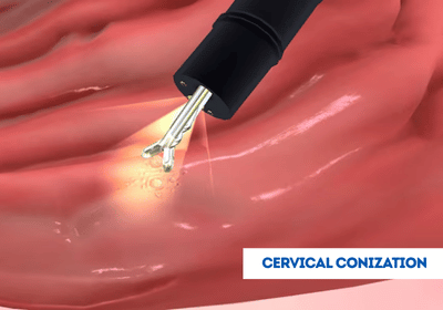
Cervical conization, or cone biopsy, is a surgical procedure in which a cone-shaped piece of cervical tissue is removed for diagnosis or treatment. It is often used to detect and manage precancerous or cancerous lesions, helping doctors plan the right care. International patients can access the cervical conization procedure in India, Turkey, Thailand, Dubai, and other leading countries at advanced hospitals with experienced specialists, coordinated by EdhaCare.
[Book Consultation & Get Treatment Quote – India | Turkey | Thailand | Dubai]
Cervical conization is a surgical procedure performed to remove a cone-shaped section of tissue from the cervix. The removed tissue includes both the outer surface of the cervix (ectocervix) and the inner canal (endocervix). This procedure allows for a closer examination of the cervical tissue and helps diagnose and treat certain cervical conditions.
The cost of cervical conization can vary based on the method used, the healthcare facility, and the location. Below are approximate estimated costs for cervical conization in India, Turkey, Thailand, and Dubai:
| Country | Approximate Cost |
|---|---|
| India | USD 300 – 1,000 |
| Turkey | USD 500 – 1,500 |
| Thailand | USD 600 – 2,000 |
| Dubai | USD 1,000 – 3,000 |
Note: These costs are approximate and may vary based on the hospital, procedure specifics, and additional treatments. Contact EdhaCare for more accurate cost of cervical conization in India.
EdhaCare helps patients access the best hospitals in India for cervical conization. We connect you with top hospitals for cervical conization in India offering advanced surgical techniques and expert care.
| Hospital Name | Location |
|---|---|
| Fortis Hospital - Shalimar Bagh | New Delhi |
| Artemis Hospital | Gurgaon |
| Apollo Hospital | Navi Mumbai |
| Manipal Hospital - Kharadi | Pune |
| CK Birla Hospital | Jaipur |
Through EdhaCare, connect with the best doctors in India for cervical conization. These top doctors for cervical conization in India provide safe procedures and international patient support.
| Doctor Name | Hospital Name | Location |
|---|---|---|
| Dr. Shakti Bhan Khanna | Apollo | Delhi |
| Dr. Savitri Subramanyam | Vijaya | Chennai |
| Dr. Jaya M Bhat | Fortis | Bengaluru |
| Dr. Anita Kant | Asian | Faridabad |
| Dr. Girish Sabnis | Narayana | Mumbai |
Cervical conization means the removal of a cone-shaped piece of tissue from the cervix. This can be done using a few different techniques:
Cone biopsy, while generally safe, carries some risks. These may include:
After a cone biopsy or cervical conization, patients enter a recovery phase that requires careful monitoring and care. The first few days may involve mild cramping and spotting, which are normal reactions to the procedure. It’s essential to rest and avoid strenuous activities for at least a week to allow the cervix to heal.
Patients are advised to refrain from sexual intercourse, using tampons, or douching for at least two to four weeks. These actions can introduce bacteria and increase the risk of infection. It’s also crucial to watch for any unusual symptoms, such as heavy bleeding, severe pain, or signs of infection like fever or foul-smelling discharge.
Follow-up appointments with the healthcare provider are necessary to ensure proper healing and discuss any further treatment if needed. Understanding these guidelines helps promote a smooth recovery and reduces potential complications during the post-conization period.
Cervical conization, although usually safe, does involve potential complications. This is the breakdown of the risks involved:
The procedure for cervical conization typically involves the following steps:
Outcomes of cervical conization can be quite variable, and it is necessary to understand what the results would mean. Given below, in short, will be possible outcomes:
Pathology Evaluation: The cone of cervical tissue is processed to a lab search for normal cells, precancerous cells (cervical dysplasia or CIN), or cancer cells.
Margin Status: The tissue margin is defined in the pathology report. Negative margins are indicative of the complete excision of abnormal tissue. Positive margins imply some abnormal tissue may remain.

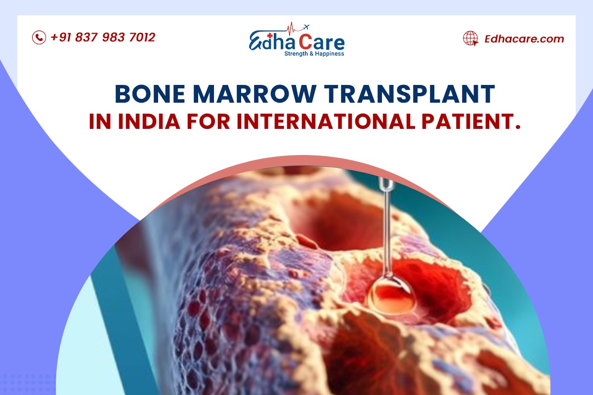
A bone marrow transplant can be a life-changing treatment for people living with serious blood cance...

Lung cancer is the most commonly diagnosed cancer across the globe and it serves as the primary reas...

Are you planning to travel from the United Kingdom to India for your treatment but uncertain about t...