

Require Assistance?
Get A Quick Callback From Our Healthcare Experts
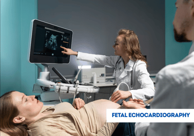
Fetal echocardiography is a specialized ultrasound procedure that examines the structure and function of a developing baby’s heart during pregnancy. It provides detailed images to detect any potential heart abnormalities and assess fetal cardiac health. International patients can access fetal echocardiography in India, Turkey, Thailand, Dubai, and other leading countries at advanced hospitals, with EdhaCare coordinating care and support throughout the process.
[Book Consultation & Get Treatment Quote – India | Turkey | Thailand | Dubai]
The primary purpose of fetal echocardiography is to assess the structure and function of the fetal heart and detect any potential abnormalities or congenital heart defects. It helps healthcare providers identify conditions such as:
By identifying these conditions before birth, healthcare providers can plan appropriate medical interventions and develop a comprehensive postnatal care plan for the baby.
The cost of fetal echocardiography varies depending on the healthcare facility, the expertise of the specialist, and the location. Below are approximate estimated costs for fetal echocardiography in India, Turkey, Thailand, and Dubai:
| Country | Approximate Cost |
|---|---|
| India | USD 100 – 400 |
| Turkey | USD 200 – 600 |
| Thailand | USD 250 – 700 |
| Dubai | USD 400 – 1,200 |
Note: These costs are approximate, For the most accurate cost of fetal echocardiography and personalized guidance, please contact EdhaCare.
EdhaCare introduces the best hospitals in India for fetal echocardiography, helping you find the top hospitals for fetal echocardiography in India equipped with advanced fetal imaging and diagnostic technology.
| Hospital Name | Location |
|---|---|
| Fortis Hospital - Shalimar Bagh | New Delhi |
| Artemis Hospital | Gurgaon |
| Apollo Hospital | Navi Mumbai |
| Manipal Hospital - Kharadi | Pune |
| CK Birla Hospital | Jaipur |
Connect with the best doctors in India for fetal echocardiography, chosen by EdhaCare for their expertise. These top doctors for fetal echocardiography in India ensure accurate diagnosis and prenatal care.
| Doctor Name | Hospital Name | Location |
|---|---|---|
| Dr. Shakti Bhan Khanna | Apollo | Delhi |
| Dr. Savitri Subramanyam | Vijaya | Chennai |
| Dr. Jaya M Bhat | Fortis | Bengaluru |
| Dr. Anita Kant | Asian | Faridabad |
| Dr. Girish Sabnis | Narayana | Mumbai |
The procedure for fetal echocardiography generally follows these steps:


Lung cancer is the most commonly diagnosed cancer across the globe and it serves as the primary reas...

Are you planning to travel from the United Kingdom to India for your treatment but uncertain about t...

Planning to travel from Germany to India for medical treatment? Confused about the steps involved in...