


Require Assistance?
Get A Quick Callback From Our Healthcare Experts
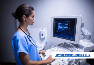
Hysterosonography, also called sonohysterography, is a minimally invasive diagnostic procedure that uses ultrasound to examine the uterus and its lining. It provides detailed insights to help doctors diagnose and monitor various gynecological conditions. With the support of EdhaCare, international patients can access advanced hysterosonography services in India, Turkey, Thailand, Dubai, and other leading countries at top hospitals with experienced specialists.
[Book Consultation & Get Treatment Quote – India | Turkey | Thailand | Dubai]
Hysterosonography involves the introduction of a sterile saline solution into the uterus, followed by the use of ultrasound imaging to visualize the uterine cavity. The saline infusion helps to expand the uterine cavity, providing a clearer image of the uterine lining and any potential abnormalities.
Hysterosonography may be performed for several reasons, including:
The specific reasons for performing hysterosonography may vary depending on the individual's symptoms and medical history.
Below are approximate estimated costs for hysterosonography in India, Turkey, Thailand, and Dubai:
| Country | Approximate Cost |
|---|---|
| India | USD 100 – 400 |
| Turkey | USD 150 – 500 |
| Thailand | USD 200 – 600 |
| Dubai | USD 400 – 1,000 |
Note: The above cost is only an estimate. Reach out to EdhaCare to know the exact hysterosonography medical procedure cost in India based on your medical requirements.
EdhaCare introduces the best hospitals in India for hysterosonography, helping you find the top hospitals for hysterosonography in India offering advanced ultrasound diagnostics and expert gynecological care.
| Hospital Name | Location |
|---|---|
| Fortis Hospital - Shalimar Bagh | New Delhi |
| Artemis Hospital | Gurgaon |
| Apollo Hospital | Navi Mumbai |
| Manipal Hospital - Kharadi | Pune |
| CK Birla Hospital | Jaipur |
Connect with the best doctors in India for hysterosonography, chosen by EdhaCare for their experience. These top doctors for hysterosonography in India ensure precise evaluation and patient safety.
| Doctor Name | Hospital Name | Location |
|---|---|---|
| Dr. Shakti Bhan Khanna | Apollo | Delhi |
| Dr. Savitri Subramanyam | Vijaya | Chennai |
| Dr. Jaya M Bhat | Fortis | Bengaluru |
| Dr. Anita Kant | Asian | Faridabad |
| Dr. Girish Sabnis | Narayana | Mumbai |
Learn about hysterosonography, a diagnostic procedure for evaluating the uterus. Discover the hysterosonography procedure and its importance in gynecology.The procedure for hysterosonography typically involves the following steps:


Lung cancer is the most commonly diagnosed cancer across the globe and it serves as the primary reas...

Are you planning to travel from the United Kingdom to India for your treatment but uncertain about t...

Planning to travel from Germany to India for medical treatment? Confused about the steps involved in...