Require Assistance?
Get A Quick Callback From Our Healthcare Experts
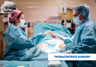
Thoracentesis treatment involves a minimally invasive procedure to remove excess fluid from the pleural cavity, often performed to diagnose or relieve conditions such as infections, heart failure, or cancer. EdhaCare assists international patients seeking thoracentesis surgery in India, Turkey, Thailand, Dubai, and other countries by connecting them with experienced doctors and NABH- or JCI-accredited hospitals, providing expert consultation, personalized treatment planning, and coordinated post-treatment care.
[Book Consultation & Get Treatment Quote – India | Turkey | Thailand | Dubai]
Thoracentesis, also known as pleural tap, is a medical procedure used to remove pleural fluid for analysis or symptom relief. It helps diagnose the cause of fluid buildup and can significantly improve breathing comfort. A needle or thin tube (catheter) is inserted into the chest cavity under local anesthesia, often guided by ultrasound or imaging. Thoracentesis Surgery in India is performed with utmost precision to minimize risks and ensure quick recovery for patients suffering from pleural effusion or related lung conditions.
The cost of thoracentesis surgery varies depending on the hospital, procedure complexity, underlying condition, and specialist expertise. Below are approximate costs for thoracentesis surgery in India, Turkey, Thailand, and Dubai:
| Country | Approximate Cost |
|---|---|
| India | USD 300 – 1,200 |
| Turkey | USD 800 – 2,000 |
| Thailand | USD 1,000 – 2,500 |
| Dubai | USD 1,500 – 3,500 |
Note: The above costs of thoracentesis surgery are only estimates. Reach out to EdhaCare for the exact thoracentesis surgery cost and personalized treatment guidance.
EdhaCare guides global patients to the best hospitals in India for thoracentesis surgery, connecting them with the top hospitals for thoracentesis surgery in India offering advanced pleural care, minimally invasive procedures, and specialized pulmonology support.
| Hospital Name | Location |
|---|---|
| Sri Ramachandra Medical Center | Chennai |
| Delhi Heart and Lung Institute | Delhi |
| Calcutta Medical Research Institute (CMRI) | Kolkata |
| Manipal Hospital | Pune |
| Fortis Hospital | New Delhi |
Doctors may recommend Thoracentesis Surgery in India for a range of medical conditions, including:
Removing and analyzing the pleural fluid helps doctors identify the underlying cause and plan further treatment effectively.
Patients may require a thoracentesis if they experience the following symptoms:
Undergoing Thoracentesis Surgery in India can relieve these symptoms quickly and improve lung function.
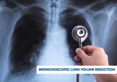
Before performing the procedure, doctors carry out several diagnostic tests to ensure accuracy and safety, such as:
These assessments help pulmonologists plan a safe and effective Thoracentesis Surgery in India.
Depending on the purpose and patient’s condition, thoracentesis can be of two main types:
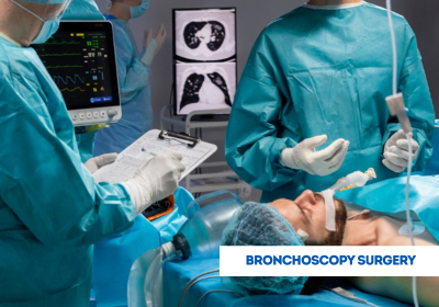
India’s advanced hospitals use cutting-edge imaging and equipment to perform both types of Thoracentesis Surgery in India safely and effectively.
During Thoracentesis Surgery in India, the patient is positioned sitting upright or slightly leaning forward. After applying local anesthesia, the doctor inserts a thin needle or catheter between the ribs into the pleural space. Fluid is then gently withdrawn and collected for testing. The entire procedure usually takes 20 to 40 minutes. Post-procedure, patients are monitored to ensure there are no complications such as infection, bleeding, or pneumothorax (air leakage). Most patients can return home the same day.
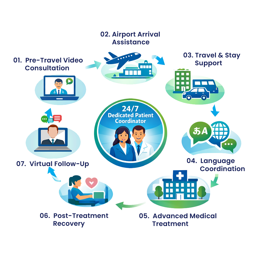
EdhaCare, a leading medical tourism company in India, connects international patients with top hospitals and respiratory specialists offering expert Thoracentesis Surgery in India. With personalized care, transparent pricing, and end-to-end medical support, EdhaCare ensures a comfortable and worry-free healthcare experience. Patients choose EdhaCare for:
With EdhaCare’s trusted network and comprehensive medical tourism package for Thoracentesis Surgery, patients receive safe, effective, and affordable Thoracentesis Surgery in India, ensuring better breathing, accurate diagnosis, and long-term lung health.


Lung cancer is the most commonly diagnosed cancer across the globe and it serves as the primary reas...

Are you planning to travel from the United Kingdom to India for your treatment but uncertain about t...

Planning to travel from Germany to India for medical treatment? Confused about the steps involved in...